Control and Coordination
📋 Table of Contents
1. Introduction — What is Control & Coordination?
The reasons that living things require a control system
Every living thing engages in a variety of life functions, including growth, digestion, reproduction, and movement. For these processes to happen in a systematic and orderly way, the body needs a control and coordination system.
DefinitionControl refers to the regulation of body activities, while Coordination is the working together of different organs and systems to produce a proper response to stimuli.
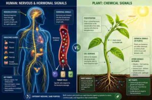
In animals, control and coordination are achieved by two systems:
- 🧠 Nervous System — fast electrical signals via neurons
- 💉 Endocrine System — slow chemical signals via hormones in blood
In the plant there are no nerves or blood. Plants use chemical control through plant hormones (phytohormones) to respond to stimuli like light, gravity, and touch.
2. Neuron — Structure and Types
A neurone (nerve cell) is the basic structural and functional unit of the nervous system. It is specialised to receive and transmit electrical impulses.
Parts of a Neuron
| Part | Description | Function |
|---|---|---|
| Cell Body (Cyton/Soma) | Contains nucleus and cytoplasm | Metabolic centre of the neuron |
| Dendrites | Short, branched extensions from cell body | Receive impulses from other neurons |
| Axon | Long single fibre; may be myelinated | Carries impulses away from cell body |
| Myelin Sheath | Fatty insulating layer around axon | Speeds up nerve impulse transmission |
| Nodes of Ranvier | Gaps in myelin sheath | Allow faster saltatory conduction |
| Synapse | Junction between two neurons | Transfer impulse via neurotransmitters |
| Axon Terminal | End knobs at tip of axon | Release neurotransmitters |
Types of Neurons
| Type | Also Called | Direction of Impulse | Function |
|---|---|---|---|
| Sensory Neuron | Afferent | Sense organ → Brain/Spinal cord | Carries sensory information |
| Motor Neuron | Efferent | Brain/Spinal cord → Muscle/gland | Carries response commands |
| Relay Neuron | Interneuron/Association | Within CNS | Connects sensory to motor neurons |
3. The Nervous System
Organisation of the human nervous system
Central Nervous System (CNS)
Peripheral Nervous System (PNS)
Spinal CordSomatic NS
Autonomic NS
| Division | Components | Function |
|---|---|---|
| CNS | Brain + Spinal Cord | Processing and integration of information |
| PNS | All nerves outside CNS | Relay signals to and from CNS |
| Somatic NS | Voluntary nerves | Controls skeletal muscles; voluntary movement |
| Autonomic NS | Sympathetic + Parasympathetic | Controls involuntary functions (heartbeat, digestion) |
Key Point
The nervous system uses electrochemical impulses . Neurones transmit electrical signals. Chemical signals (neurotransmitters such as acetylcholine) cross the synaptic cleft between neurones.
4. Reflex Action and Reflex Arc
Reflexes Involuntary, spontaneous reactions to stimuli
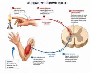
DefinitionA reflex action is a sudden, involuntary, and automatic response to a stimulus. It does NOT require thinking. For example, withdraw hand when touching a hot object.
Reflex Arc — Pathway of a Reflex
→
Receptor
→
Sensory Neuron
→
Spinal Cord (Relay)
←
Muscle / Gland
←
Motor Neuron
| Component | Role | Example |
|---|---|---|
| Receptor | Detects stimulus | Skin receptors (pain, heat) |
| Sensory Neuron | Carries signal to spinal cord | Afferent nerve fibre |
| Relay Neuron | Processes in spinal cord | Interneuron in grey matter |
| Motor Neuron | Carries command to effector | Efferent nerve fibre |
| Effector | Produces response | Muscles, glands |
Important Exam PointReflex arcs are controlled by the spinal cord, NOT the brain. The brain is informed about the reflex after the response has occurred. This makes reflexes faster and saves time in emergencies.
5. Human Brain — Structure and Functions
The master control centre of the body
The human brain is protected by the skull (cranium) and surrounded by three membranes called meninges. Between the meninges is cerebrospinal fluid (CSF) which acts as a shock absorber.
| Part | Sub-parts | Key Functions |
|---|---|---|
| Forebrain Front | Thalamus, Cerebrum, Hypothalamus | Thought, memory, intelligence, temperature and hunger regulation, sensory relay |
| Midbrain, middle | Superior and inferior colliculi | Visual reflexes and auditory reflexes; balance |
| Hindbrain (Rear) | Medulla oblongata, Pons, and Cerebellum | Balance & posture. Breathing bridge. Controls heartbeat/breathing/BP (vital centres). |
Functions of Cerebrum Lobes
| Lobe | Location | Function |
|---|---|---|
| Frontal Lobe | Front | Thinking, planning, speech (Broca’s area), personality |
| Parietal Lobe | Top-rear | Sensory perception, spatial awareness, touch |
| Temporal Lobe | Sides | Hearing, language understanding (Wernicke’s area), memory |
| Occipital Lobe | Back | Vision and visual processing |
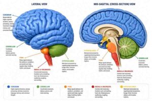
Memory TrickMedulla = Maintains Life (controls heartbeat, breathing, blood pressure — the involuntary vital functions we cannot survive without)
6. How Does the Nervous System Work?
Impulse transmission and synaptic signalling
Steps of Nerve Impulse Transmission
- A stimulus (heat, light, sound, touch) is detected by receptor cells.
- The receptor converts stimulus to an electrical impulse.
- The impulse travels along the sensory neuron to the CNS.
- At the synapse, the electrical impulse triggers release of chemical neurotransmitters.
- Neurotransmitters diffuse across the synaptic cleft and bind to receptors on the next neuron.
- A new impulse is generated in the next neuron.
- The signal reaches the effector (muscle or gland) via motor neuron.
- The effector produces a response.
How Impulse Travels in Nerve Cell
- Resting neuron: inside is negatively charged (more K⁺ inside, more Na⁺ outside)
- During impulse: Na⁺ ions rush in → inside becomes positively charged (depolarisation)
- After impulse: K⁺ ions rush out → membrane repolarises
- This wave of electrochemical change = action potential
7. Chemical Coordination — Endocrine System & Hormones
Slow but far-reaching chemical messengers
DefinitionHormones are chemical messengers secreted directly into the bloodstream by ductless glands (endocrine glands) to regulate body functions at distant target organs.
Major Endocrine Glands and Their Hormones
| Gland | Location | Hormone(s) | Function | Deficiency Disease |
|---|---|---|---|---|
| Pituitary Master | Base of brain | GH, TSH, FSH, LH, ADH, Oxytocin | Controls all other glands; growth, water balance, reproduction | Dwarfism (GH↓) / Gigantism (GH↑) |
| Thyroid | Neck (front) | Thyroxine | Regulates metabolism, growth, development | Goitre, Cretinism |
| Parathyroid | Behind thyroid | Parathormone (PTH) | Regulates calcium & phosphorus levels | Tetany |
| Adrenal | Above kidneys | Adrenaline (Epinephrine), Cortisol | Fight-or-flight response; stress hormones; salt balance | Addison’s disease |
| Pancreas Dual | Abdomen | Insulin, Glucagon | Regulates blood sugar (glucose) | Diabetes mellitus (Insulin↓) |
| Testes | Scrotum (male) | Testosterone | Male secondary sexual characters; sperm production | Delayed puberty |
| Ovaries | Abdomen (female) | Oestrogen, Progesterone | Female sexual characters; menstrual cycle; pregnancy | Irregular menstruation |
| Pineal | Brain (epithalamus) | Melatonin | Regulates sleep-wake cycle (circadian rhythm) | Sleep disorders |
Adrenaline — The Emergency Hormone
Adrenaline (also called epinephrine) is secreted by the adrenal medulla in response to stress or danger. It gets the body ready for the fight-or-flight reaction:
- ⬆️ Heart rate increases → more blood to muscles
- ⬆️ Breathing rate increases → more oxygen supply
- ⬆️ Blood sugar rises → more energy available
- ↔️ Blood diverted from digestion to muscles and brain
- 👁️ Pupils dilate → better vision in emergencies
Iodine & Thyroxine — Exam FavouriteThyroxine requires iodine for its synthesis. Deficiency of iodine → reduced thyroxine → pituitary secretes more TSH → thyroid gland enlarges → Goitre. Prevention: use iodised salt.
8. Control and Coordination in Plants
Plant movements and phytohormones
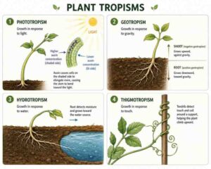
Plants do not have a nervous system. They respond to stimuli using chemical signals (plant hormones / phytohormones) and show two types of movements: Tropic movements (directional, growth-based) and Nastic movements (non-directional).
Types of Plant Movements (Tropism)
| Type | Stimulus | Direction of Growth | Example |
|---|---|---|---|
| Phototropism | Light | Shoot → towards light (+ve); Root → away (-ve) | Sunflower facing sun |
| Geotropism | Gravity | Root → towards gravity (+ve); Shoot → away (-ve) | Roots growing downward |
| Hydrotropism | Water | Roots → towards water (+ve) | Root bends toward moisture |
| Chemotropism | Chemical | Towards/away from chemical | Pollen tube grows toward ovule |
| Thigmotropism | Touch | Along support surface | Tendril of pea plant curling |
| Nastic Movement | Touch/light | Non-directional | Mimosa pudica (touch-me-not) |
“Four panels showing plant tropisms: 1) Phototropism – seedling bending toward light source with auxin distribution arrows; 2) Geotropism – root growing downward, shoot growing up; 3) Hydrotropism – root curving toward water; 4) Thigmotropism – pea tendril coiling around a support. Botanical illustration style, green and yellow tones.”
Plant Hormones (Phytohormones)
| Hormone | Type | Produced In | Main Function |
|---|---|---|---|
| Auxin (IAA) | Promoter | Shoot tips | Cell elongation; phototropism; apical dominance; prevents leaf fall |
| Gibberellin | Promoter | Young leaves, seeds | Stem elongation; seed germination; fruit development; breaks dormancy |
| Cytokinin | Promoter | Roots, developing fruits | Cell division; delays ageing (senescence); promotes lateral bud growth |
| Abscisic Acid (ABA) | Inhibitor | Leaves, stems, roots | Stomata closure (water stress); seed dormancy; promotes ageing and abscission |
| Ethylene | Gas | Ripening fruits | Fruit ripening; promotes senescence; inhibits stem elongation |
How Auxin Causes Phototropism
- Light falls on shoot tip from one side
- Auxin migrates to the shaded (dark) side
- Higher auxin concentration → more cell elongation on shaded side
- Shoot bends toward light ✔
9. Quick Comparison Tables
Nervous vs Hormonal | Plants vs Animals | Voluntary vs Involuntary
Nervous System vs Endocrine System
| Feature | Nervous System | Endocrine System |
|---|---|---|
| Signal type | Electrical impulse | Chemical (hormones) |
| Speed | Very fast (milliseconds) | Slow (seconds to days) |
| Duration | Short-lived | Long-lasting |
| Transmission medium | Nerve fibres | Blood/lymph |
| Target | Specific organ | Widespread target organs |
| Examples | Reflex, voluntary movement | Puberty, blood sugar control |
Voluntary vs Reflex Action
| Feature | Voluntary Action | Reflex Action |
|---|---|---|
| Control | Brain (cerebrum) | Spinal cord |
| Speed | Slower | Very fast |
| Consciousness | Conscious, wilful | Unconscious, automatic |
| Examples | Writing, dancing | Knee-jerk, eye blinking |
Plant Hormones — Summary Mnemonics
| Remember | Hormone | Key Word |
|---|---|---|
| A | Auxin | Apical dominance, Accelerates cell elongation |
| G | Gibberellin | Germination, Growth of stem |
| C | Cytokinin | Cell division, Counters ageing |
| A | Abscisic Acid | Abscission, Anti-growth, dormAncy |
| E | Ethylene | End of ripening, sEnescence |
“Side-by-side infographic comparing nervous system vs endocrine system: left panel shows a human brain with electric bolt symbols and fast arrows; right panel shows hormone molecules floating in bloodstream. Use blue vs amber colour coding. Include speed indicator and signal type labels. Modern flat design.”
Most Asked Exam Questions — Control & Coordination
- Draw and label the structure of a neuron.
- Explain reflex arc with a diagram.
- What is the role of the cerebellum and medulla?
- Distinguish between tropic and nastic movements.
- Why is iodine necessary for thyroid function?
- What happens in the body when adrenaline is secreted?
- How does auxin cause phototropism in plants?